What is IntelliStudio?
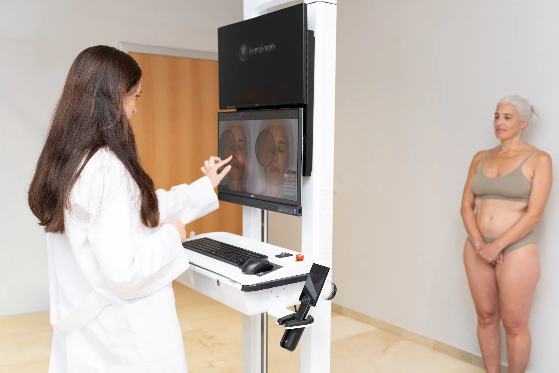
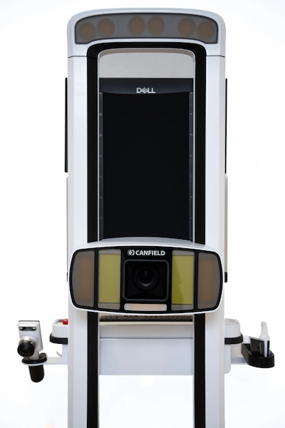
IntelliStudio by Canfield is an advanced clinical photography system that allows specialists to analyze, document, and accurately monitor the skin of the face and body over time. Thanks to its high-quality images and standardized image capture, specialists at Clínica Planas can examine specific skin characteristics and compare changes between appointments.
Its applications include:
- Dermatological monitoring of moles and other skin lesions.
- Assessment of pigmentation, redness, and signs of photoaging.
- Monitoring of dermatological and aesthetic treatments.
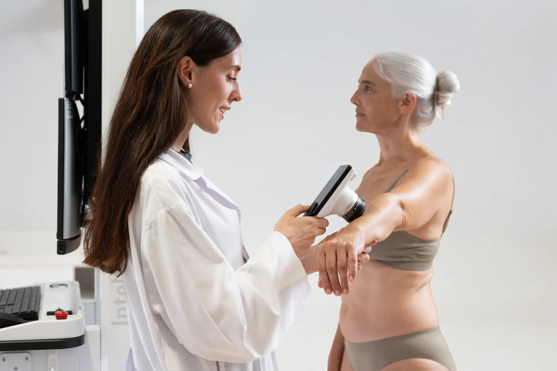
At Clínica Planas, IntelliStudio is complemented by Canfield's D2 digital dermatoscope, which allows specialists to capture magnified images of selected skin lesions and the scalp.
More than simply taking photographs of your skin, it is a tool that helps specialists understand how your skin changes over time and make better-informed medical decisions. IntelliStudio does not treat skin conditions itself, nor does it replace a medical examination, dermoscopy interpreted by a specialist, or any additional tests that may be necessary.
Book an
appointment
CLÍNICA PLANAS' DATA PROTECTION INFORMATION CLICK HERE
Purposes: To respond to your requests and to send you commercial information about our products and services, including via electronic means. Rights: At any time, you can withdraw your consent, access, rectify, delete your data and exercise other rights at doctor@clinicaplanas.com. Additional Information: Privacy Policy.

Our Patients’
Reviews
 Raquel Pacheco
12/08/2026

 Robbie Berman
11/08/2026
 M.Claustre Besora Canal
02/07/2026
 Cristina López sole
09/06/2026
Features of the IntelliStudio system
The main advantage of IntelliStudio is its ability to capture comparable images at different points in time. Positioning, framing, and lighting are standardized so that specialists can assess changes more reliably than with conventional photographs.
MatchPose technology
MatchPose helps reproduce the positioning used in previous photographs. This means that when you return for a follow-up appointment, the same area can be photographed from similar angles, allowing specialists to monitor changes in moles, pigmentation, skin lesions, or treatment results with greater accuracy.
Intelligent facial detection
The system recognizes facial reference points to facilitate consistent and reproducible facial photography. This standardization is particularly useful when comparing images of pigmentation, redness, wrinkles, and other skin characteristics across different appointments.
Guided and automated image capture
During the session, a healthcare professional will guide you on how to position yourself in front of the camera. The system uses predefined positions and framing and automates part of the image capture process, making it easier to document different areas of the body under similar conditions. Each session is adapted to the areas your doctor needs to examine.
Integrated video
IntelliStudio also includes video capabilities that can complement photographic documentation of specific areas. This feature provides additional perspectives when the specialist considers that a still image is insufficient to document an area of interest.
Lighting and visualization modes
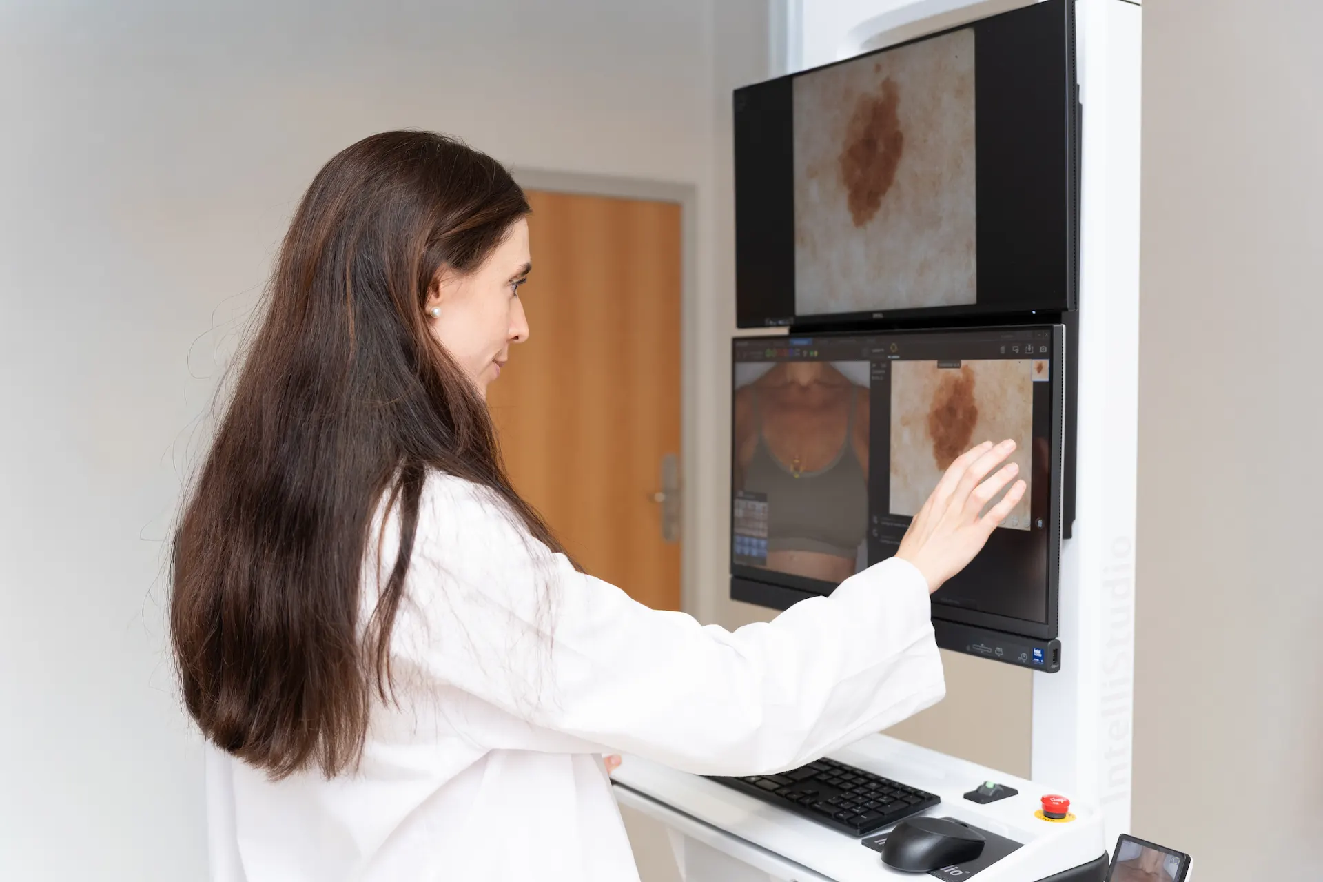
Not all skin characteristics are visible under the same lighting conditions. IntelliStudio offers different image capture and visualization modes that can provide additional information about pigmentation, redness, and other skin concerns.
Polarized light
Polarized light reduces reflections on the skin's surface, making it easier to observe certain skin structures and characteristics. It can be useful for documenting pigmentation, redness, and vascular irregularities and comparing their progression over time, always as a complement to a dermatological assessment.
RBX
RBX is an image-processing technology developed by Canfield that highlights components related to skin pigmentation and vascularization. It can provide additional visual information when assessing and monitoring conditions such as melasma, sunspots, and certain types of redness.
Applications of the system
IntelliStudio can be used in clinical and aesthetic dermatology to document the initial condition of the skin, assess changes, and make visual comparisons between appointments. Its usefulness depends on the medical indication and the areas that need to be documented.
Before-and-after photography
Photographs taken under similar conditions allow specialists to compare the appearance of the skin over time. In dermatology, they facilitate the monitoring of moles and skin lesions, particularly in patients with numerous nevi, a history of melanoma, or other risk factors identified by the specialist.
When indicated, dermatologists can complement full-body photographs with D2 digital dermoscopy of specific lesions. The frequency of follow-up examinations is determined individually; in some cases, annual or six-monthly check-ups may be recommended. In aesthetic medicine, photographic documentation also helps assess treatment progress and maintain realistic expectations.
Facial assessment
Did you know that some skin conditions can be difficult to detect with the naked eye? IntelliStudio images complement facial examinations and allow specialists to document pigmentation, redness, vascular characteristics, and signs of photoaging.
Based on this information, the specialist can provide personalized skincare recommendations and, when appropriate, suggest dermatological or aesthetic treatments suited to your skin. Image interpretation and diagnosis always remain the responsibility of the doctor.
Pigmentation assessment
Skin pigmentation can be associated with sun exposure, hormonal factors, or inflammatory processes, among other causes. IntelliStudio helps document the distribution of pigmentation and compare changes over time. Its visualization modes can provide additional information when monitoring melasma, solar lentigines, and other pigmentation disorders.
The aim is to create a visual record that helps assess the skin's response to recommended skincare and treatments, rather than independently predicting the appearance of new pigmentation.
Hair Unit
Have you noticed that your hair has lost density, become thinner, or is shedding more than usual? Understanding the condition of your scalp and monitoring changes over time is important for assessing possible causes and ensuring appropriate follow-up care.
At the Hair Unit of Clínica Planas, IntelliStudio clinical photography can be combined with the D2 digital dermatoscope to capture detailed images of the scalp and hair. Through digital trichoscopy, specialists can examine specific hair characteristics and complement their clinical assessment.
This documentation can be useful in cases of:
- Androgenetic alopecia.
- Telogen effluvium.
- Alopecia areata.
- Other scalp conditions.
It allows specialists to document the initial condition and compare photographs taken during subsequent appointments to assess changes in hair density, appearance, and the areas being examined.
It can also be used to document progress during medical treatments for alopecia, growth factor therapies, mesotherapy, certain laser or LED procedures, and, when appropriate, before and after hair transplantation. These images help monitor treatment response but do not replace a medical diagnosis or guarantee specific results.
Treatments associated with IntelliStudio
IntelliStudio does not perform aesthetic or dermatological treatments itself. Instead, it allows specialists to document the condition of the skin and compare changes before, during, and after certain procedures when considered medically useful.
Dermatological monitoring of moles and skin lesions
Standardized body photography allows specialists to document the distribution and appearance of moles and other skin lesions. By comparing images from different appointments, dermatologists can identify changes that may require closer examination and determine whether additional tests are necessary.
Full-body mole mapping (body mapping)
When numerous moles or lesions are distributed across different areas of the body, body mapping helps create an organized visual record. Photographs taken from multiple positions serve as references for future examinations and make it easier to locate specific lesions.
Dermatological monitoring for patients at risk of skin cancer
For patients with a personal or family history of melanoma, atypical moles, or other risk factors, specialists may recommend regular photographic monitoring. IntelliStudio complements dermatological examinations and helps document changes, but it cannot independently diagnose skin cancer or replace a biopsy when one is indicated.
Digital dermoscopy of moles and pigmented lesions
The D2 digital dermatoscope, used alongside IntelliStudio, allows specialists to capture magnified images of lesions selected by the dermatologist. These images provide additional information about their structure and can be compared during subsequent check-ups to support medical decision-making.
Monitoring new or changing skin lesions
When a new skin lesion appears or an existing lesion changes in appearance, the dermatologist may decide to document it using clinical photography and, when appropriate, digital dermoscopy. This record facilitates comparisons between appointments. Any suspicious lesion requires medical assessment without waiting for the next scheduled photographic examination.
Monitoring psoriasis, vitiligo, and other skin conditions
In certain widespread skin conditions, such as psoriasis or vitiligo, standardized photographs can help document affected areas and monitor their progression. Their usefulness depends on each individual case and the dermatologist's assessment.
Monitoring scars and dermatological surgery
Images captured before and after a dermatological procedure allow specialists to document the appearance of the treated area and monitor its progression. They can also be useful for tracking certain scars, always as a complement to medical follow-up.
Treatments for facial pigmentation and dark spots
In cases of melasma, solar lentigines, or other pigmentation disorders, clinical photography allows specialists to document the skin's initial condition and compare changes over time. The available lighting and visualization modes can provide additional information to support the specialist's assessment.
Dermatological and facial laser treatments
Photographs taken under similar conditions allow specialists to document the progress of certain laser treatments, both for pigmented lesions and other dermatological indications. The assessment of results remains the responsibility of the treating professional and depends on the procedure performed.
Monitoring rosacea, redness, and vascular conditions
For patients with rosacea, erythema, or small visible dilated blood vessels, photographic documentation can help compare the appearance of redness across different appointments. This monitoring provides an additional visual reference for assessing the response to recommended skincare or treatments.
Assessment of photoaging and sun damage
IntelliStudio allows specialists to document visible characteristics associated with sun exposure and skin aging, such as changes in pigmentation or texture. Comparing images helps monitor the skin's progression and provides context for the specialist's recommendations.
Facial rejuvenation and wrinkle treatments
For procedures designed to improve wrinkles and expression lines, standardized photographs make it easier to compare facial appearance before and after treatment. This helps specialists observe gradual changes and maintain realistic expectations without replacing clinical assessment.
Monitoring neuromodulator treatments
Facial images can document the appearance of expression lines before and after neuromodulator treatment. Specialists can compare areas such as the forehead or eye contour, taking into account image capture conditions and individual progression.
Hair assessment and digital scalp trichoscopy
Combining clinical photography with the D2 digital dermatoscope allows specialists to obtain both general and magnified images of the scalp. Trichoscopy complements the medical examination and can help specialists assess hair and scalp conditions.
Monitoring androgenetic alopecia
In women and men with androgenetic alopecia, photographs taken during different appointments allow specialists to document hair distribution and visible changes in density. Photographic monitoring provides an objective reference that complements the specialist's assessment.
Monitoring telogen effluvium and diffuse hair loss
When diffuse hair loss occurs, specialists can use photography and trichoscopy to document the initial condition and monitor its progression. These images help provide context for changes, although the underlying cause of hair loss must be determined through an appropriate medical assessment.
Monitoring alopecia areata and scarring alopecia
In selected cases of alopecia areata or scarring alopecia, photographic documentation of affected areas can facilitate monitoring. The dermatologist will determine which areas should be documented and how frequently they should be reviewed.
Assessment of inflammatory scalp conditions
Magnified images can be useful for documenting certain inflammatory scalp conditions and comparing their progression. Diagnosis and treatment selection depend on the specialist's examination and, when appropriate, additional tests.
Monitoring medical treatments for hair loss
Standardized photography and trichoscopy allow specialists to compare hair condition during medical treatments prescribed for different forms of alopecia. The aim is to monitor progress and provide additional information during follow-up appointments.
Monitoring growth factor therapies, mesotherapy, and hair treatments
For hair treatments such as growth factor therapies, mesotherapy, or certain laser or LED light procedures, images can help document changes in hair condition. The suitability and effectiveness of each treatment must be assessed individually by the specialist.
Monitoring before and after hair transplantation
When included in the patient's treatment plan, photographic documentation allows specialists to record the condition before a hair transplant and monitor subsequent progress. Photographs taken under comparable conditions make it easier to assess changes over time.