Introduction
Oral and maxillofacial surgery is a surgical speciality encompassing the diagnosis and treatment of functional and aesthetic disorders affecting the mouth, face, neck, and craniofacial structures. The scope of this speciality encompasses reconstructive, corrective, and aesthetic facial surgery.
Book an
appointment
CLÍNICA PLANAS' DATA PROTECTION INFORMATION CLICK HERE
Purposes: To respond to your requests and to send you commercial information about our products and services, including via electronic means. Rights: At any time, you can withdraw your consent, access, rectify, delete your data and exercise other rights at doctor@clinicaplanas.com. Additional Information: Privacy Policy.

What is maxillofacial surgery?
Maxillofacial surgery is a surgical speciality involving the diagnosis, management, and rehabilitation of pathologies affecting the craniofacial region, oral cavity, and neck. The broad clinical scope of this speciality covers oral and skeletal-facial interventions to restorative plastic surgery following trauma or illness, and cosmetic procedures such as rhinoplasty, facelifts or the placement of facial prostheses or implants.
Reflecting a broad clinical versatility, the very name of this speciality, oral and maxillofacial surgery, encompasses surgical treatments within the oral cavity and complex operations involving the facial and cranial architecture.
An overview of maxillofacial surgery
At Clínica Planas, we treat a wide range of functional, aesthetic and reconstructive conditions affecting the face, oral cavity, facial bones and neck. Advanced medical and surgical techniques are integrated to provide personalised solutions in:
Facial plastic and aesthetic surgery
We perform procedures designed to improve facial harmony and proportion, such as:
- Genioplasty (chin reshaping)
- Placement of customised facial implants (in the cheekbones or mandibular angle)
- Aesthetic or reconstructive rhinoplasty
- Facelift
- Lip lift (upper lip lift)
- Facial lipofilling
- Facial feminisation or masculinisation surgery
- Orthognathic surgery
These advanced techniques address facial disproportion, age-related concerns, or dissatisfaction with particular features.
Reconstructive surgery
We specialise in correcting facial trauma, congenital anomalies, and visible scarring through sophisticated bone reshaping, facial lipofilling (autologous fat grafts), and scar correction. These restorative treatments are further enhanced by regenerative medicine techniques to achieve harmony in both facial function and aesthetics.
Our department is a centre of excellence for the surgical correction of facial paralysis and the complex transition of early facial nerve reinnervation.
Forehead lift and frontal bone reshaping and scalp advancement
This procedure is frequently indicated for female and transgender patients seeking to address a high or particularly prominent forehead. Frontal bone reshaping and scalp advancement offer a way to redefine the hairline and balance the facial thirds, improving the overall sense of natural harmony.
The bony contour above the eyebrows or the supraorbital area can also be reshaped.
Treatments for dentofacial anomalies
Our surgical protocols correct facial asymmetries, protruding or receding jaws, and functional problems with chewing, speaking or breathing. These treatments ensure your teeth align correctly while creating a more balanced, symmetrical facial profile. They are also known as orthognathic surgery.
Oral surgery
Our clinical expertise extends from intricate extractions (including wisdom teeth) and dental implantology to the surgical management of cysts, benign tumours and assorted oral lesions. We also offer pre-prosthetic surgery and bone regeneration. As pioneers in customised subperiosteal implants, we provide advanced solutions for full oral rehabilitation.
Each treatment is planned on an individual basis, incorporating 3D diagnostic imaging technologies and virtual models to ensure maximum surgical precision and optimal functional and aesthetic results.
When is maxillofacial surgery necessary?
Maxillofacial surgery is recommended when there is:
- A functional problem (difficulty chewing, biting, breathing or speaking).
- A structural problem (bone asymmetries, trauma, defects or atrophy of the jaws)
- An aesthetic requirement (improving facial contours, correcting disproportions, rejuvenation).
Each case is assessed on an individual basis to determine the most appropriate surgical approach.
What does maxillofacial surgery involve?
Depending on the diagnosis, various types of procedures may be performed, affecting both:
- Facial bone structures (jaws, mandible, chin, cheekbones, forehead, nose).
- Soft tissues (lips, cheeks, facial skin, eyelids).
- Oral cavity (gums, teeth, mucous membranes, glands).
Surgery may be reconstructive, aesthetic or functional in nature, and in many cases combines advanced surgical techniques with technologies such as 3D scanners, customised cutting guides or personalised prosthesis printing.
Related treatments
-

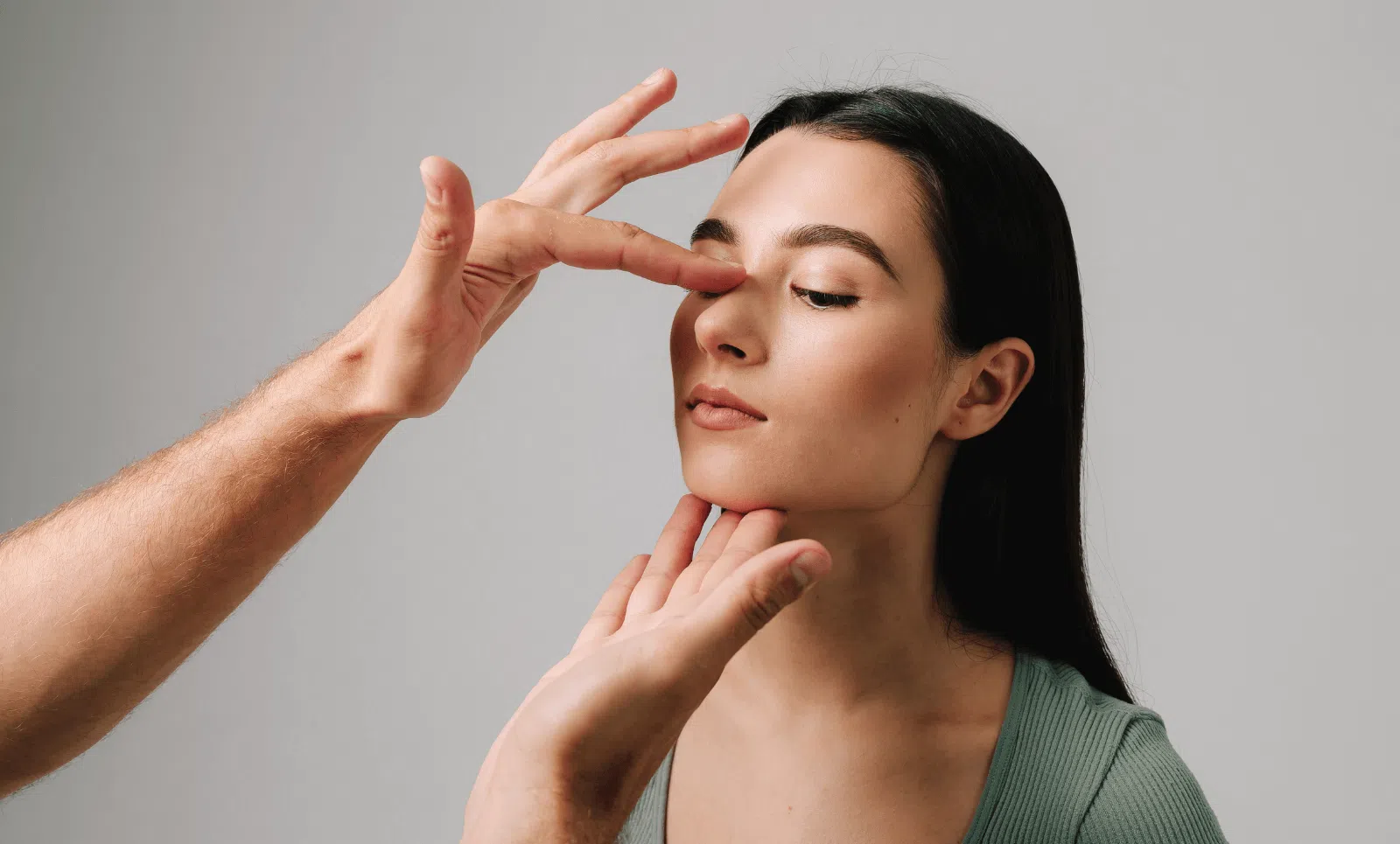
Rhinoplasty
Rhinoplasty is a surgical procedure designed to correct nasal deformities in order to improve facial balance and harmony. -

Cheek Surgery
Cheek implants help to harmonize facial features, achieving an essential balance for beauty of the face -
Chin Surgery
Chin Implants help to harmonize facial features, achieving an essential balance for beauty of the face.
Related
articles
-
¿En qué consiste la reconstrucción del tabique nasal?
La reconstrucción del tabique nasal es un procedimiento complejo y requerir varias intervenciones para reparar las estructuras destruidas. -
Opiniones sobre rinoplastia en Clínica Planas: experiencias y resultados en Barcelona
¿Buscas el mejor cirujano de nariz? Lee reseñas de Clínica Planas en Barcelona. Expertos en rinoplastia con excelentes opiniones y resultados naturales. -
Diferencias entre rinoplastia y septoplastia
En este post hablamos sobre las diferencias entre rinoplastia y septoplastia, dos cirugías de la nariz con objetivos diferentes.
Types of maxillofacial surgery
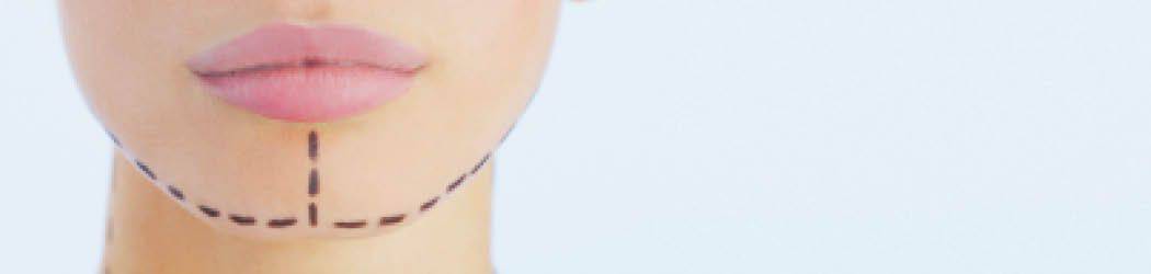
Genioplasty (mentoplasty)
Genioplasty is a surgical procedure used to reshape or adjust the position of your chin, helping to bring your facial features into a natural balance. This procedure is performed to:
- Increase projection using customised osteotomies and personalised titanium plates.
- Reduce the size of the chin following rhinoplasty, particularly in women, to achieve a more harmonious profile.
Advanced 3D scanning maps the face, ensuring every surgery is planned with maximum precision.
Mandibular angle and cheekbone implants
We use implants designed specifically for you, made from safe, biocompatible materials like PEEK, to gently enhance and define your natural facial features. They are stable, safe and removable, although they generally do not require replacement.
Forehead Reshaping and Scalp Advancement
Forehead reshaping and scalp advancement surgery. Particularly recommended for women or transgender patients with very broad foreheads or angular shapes. An incision is made along the hairline, the frontal bone is reshaped using a piezoelectric scalpel, and the scalp is advanced, visually reducing the size of the forehead and harmonising the facial proportions.
F.A.Q.
-
How much does maxillofacial surgery cost?
Every patient is unique, so costs depend on your specific procedure, the complexity of your care, any personalised materials like custom-made prostheses, and any additional tests required to ensure the best results. A personalised assessment is always carried out during a consultation to provide a bespoke quote.
-
How long does it take to recover from maxillofacial surgery?
It depends on the procedure performed, but generally, the patient can resume their normal activities within one to two weeks.
-
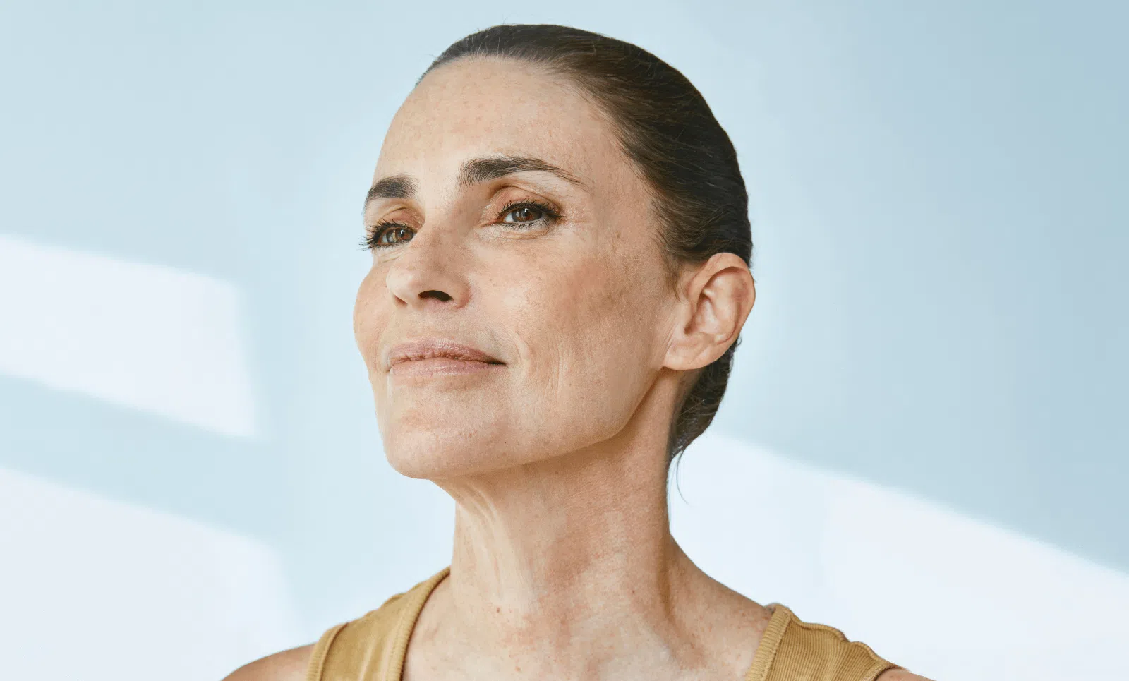
What does a person look like after maxillofacial surgery?
The main aim is to resolve the functional, aesthetic or structural problem that brought the patient to us. Through personalised planning, specialist techniques and the use of cutting-edge technology, our results are usually highly satisfactory, with a significant improvement in facial harmony, oral function and patient self-esteem.